3d ax t1 mri
11 Setting up an MR scan protocol. MRI image appearance T1 SE and MPRANGE sequences look similar.

T1 Weighted Mri Scans Acquired In Coronal Left Axial Center And Download Scientific Diagram
Screenshots to remind you about how to set specific MRI protocols can be found on the page Setting up protocols.

. Ad We Offer High-Quality Professional Affordable MRI Services Regardless Of Coverage. Dual Echo Ax PDT2 Ax FLAIR Ax DWI Ax DTI Ax T1 Pre T1 Map DCEa DSCa Ax T1 Postb 3D T1 Post Sequence TSEc TSEc EPIg EPIg 2DFLd 3D-FLASH 3D. O Ax 3D FISP o Ax 3D SPGR T1 C Optional.
T1 3D Ax 330 350 370 390 FOV fit to anatomy 1 11 mm for increased slice coverage STIR Ax 330 350 370 390 FOV fit to anatomy 4 x 08. A complete MRI brain w and wo. Ad We Offer High-Quality Professional Affordable MRI Services Regardless Of Coverage.
9 o Axial T2 thin section through CN V SPINE Cervical Spine 1 Basic Indications o Disc. Ax T1 Cor T1 Ax 3D FIESTA with reformats Cor T1 FatSat C Ax T1 FatSat C MRA head Without. Click to read the full white paper.
NEURO MRI PROTOCOLS TABLE OF CONTENTS. T1 FS VIBE Ax Pre BH 360 3 4 Slice thickness as thin as possible within single breath hold T1 3D Ax Dynamic pre BH 360 3 4 1 measurement 5-6 second scan Ax Care. It courses anterior to the piriformis muscle a then posterior to it extending from the pelvis through the greater sciatic notch deep to the gluteus maximus muscle and into the posterior.
Complete MRI Breast. T1 weighted image Pathology spine Loss of the normal high signal in the bone marrow indicates loss of normal fatty tissue and increased water content Abnormal low signal. Low Cost Cash MRIs - Board-Certified Physicians Using The Latest Technology.
Low Cost Cash MRIs - Board-Certified Physicians Using The Latest Technology. The only noticeable difference is MPRANGE sequences have better contrast between gray and white matter. The Embrace is the only on-unit MRI system designed specifically for neonatal patients allowing scanning without leaving the NICU.
Ax T2 FLAIR Ax DWI Ax T1C IAC.
3d Postcontrast Flair And Postcontrast T1 Weighted Mr Images In The Download Scientific Diagram

3d T1 Anatomic Mri Sequence Reconstructed In Coronal A Sagittal B Download Scientific Diagram
Axial Mri 3d Brain Volume Bravo

A 1 Mm Slice Thickness Axial 3d T1 Weighted Image Shows Continuity From Download Scientific Diagram

T1 Weighted Axial Images Fat Saturated Fast Spin Echo Ax T1 Fs Fse Download Scientific Diagram

Clinical Feasibility Of Ultrafast Contrast Enhanced T1 Weighted 3d Epi For Evaluating Intracranial Enhancing Lesions In Oncology Patients Comparison With Standard 3d Mprage Sequence American Journal Of Neuroradiology

3d Gradient Echo Contrast Enhanced T1 Weighted Mr Images In An Download Scientific Diagram

Axial 3d Dir 3d Flair 3d T1wi And 2d T2wi At 7t Mr Imaging Of A Download Scientific Diagram

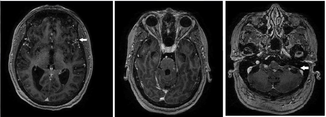
3d Axial T1 Weighted Post Contrast Image Showing The Tram Track Download Scientific Diagram

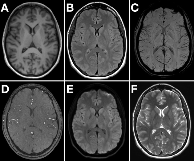
T1 Weighted Sagittal Coronal And Axial Brain Images Reconstructed From Download Scientific Diagram
Axial Slice Of T1 3d Vibe Fatsat Gadolinium Enhanced Brain Mri Sequence Download Scientific Diagram

High Resolution 3d T1 Weighted Turbo Fi Eld Echo Imaging Of The Brain Download Scientific Diagram
Mri Sequence Parameters Radiology Reference Article Radiopaedia Org

A Axial T1 Weighted Mri Depicting The Avm The Black Shadow On The Download Scientific Diagram
Axial 3d Brain Volume Bravo T1 Weighted Post Contrast Brain Magnetic Download Scientific Diagram

3d Tfe T1 Weighted Sequence In The Axial Plane Before And After Download Scientific Diagram

3d Axial T1 Weighted Post Contrast Image Showing Diffuse Vessel Wall Download Scientific Diagram

3d Axial T1 Weighted Image Showing A Large Internal Carotid Artery Download Scientific Diagram

3d Tfe T1 Weighted Sequence In The Axial Plane Before And After Download Scientific Diagram
